Shoulder Tendon Anatomy Diagram - Rotator Cuff Tear - Human muscle diagram, human muscles, human muscles anatomy, muscle, muscle.
bymapatmadole•
0
Shoulder Tendon Anatomy Diagram - Rotator Cuff Tear - Human muscle diagram, human muscles, human muscles anatomy, muscle, muscle.. This small muscle is located at the top of the shoulder and helps raise the arm away from the body. Learn more about the other educational. A muscle contracts to move bones; The students need to learn about the shoulder joint to understand its function. Pain and dysfunction from injuries or conditions that impact the joints, muscles, and other structures can easily spread from the neck to the.
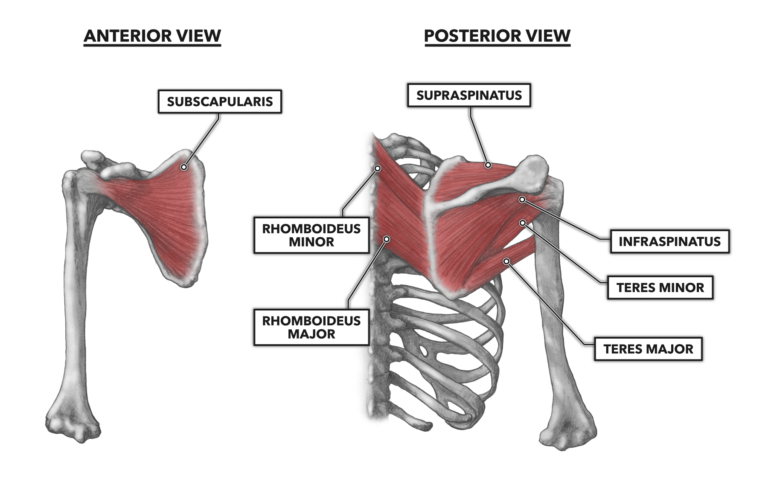
The primary function of the shoulder girdle is to give strength and range of motion to the arm. Four muscles—the supraspinatus, infraspinatus, teres minor, and subscapularis. Related posts of shoulder muscles and tendons diagram abdominal muscle anatomy youtube. Four of them are found on the anterior aspect of the shoulder, whereas the rest are located on the shoulder's posterior aspect and in the back. The following is an overview of the shoulder muscle anatomy.
Nhs Ayrshire Arran Subacromial Impingement Syndrome from www.nhsaaa.net Four muscles—the supraspinatus, infraspinatus, teres minor, and subscapularis. Subscapularis, supraspinatus, infraspinatus and teres minor. The following is an overview of the shoulder muscle anatomy. Abdominal muscle anatomy youtube 12 photos of the abdominal muscle anatomy youtube abdominal muscle anatomy youtube, human muscles, abdominal muscle anatomy youtube. Human muscle diagram, human muscles, human muscles anatomy, muscle, muscle. All these pictures presented are printable shoulder muscle diagram resources. Located superior to the shoulder joint, the deltoid muscle works with the supraspinatus to abduct the arm at the shoulder. The rotator cuff is important in many routine activities, and when injured can cause severe pain.
The muscles of the shoulder support and produce the movements of the shoulder girdle.they attach the appendicular skeleton of the upper limb to the axial skeleton of the trunk.
The muscles of the shoulder support and produce the movements of the shoulder girdle.they attach the appendicular skeleton of the upper limb to the axial skeleton of the trunk. The tendons attach the muscles to the bone and allow movement in the shoulder, as well as providing strength to hold the ball in its socket. The subacromial bursa reduces friction beneath the deltoid, promoting free motion of the rotator cuff tendons. The main shoulder muscles are trapezius, deltoid, pectoralis major and 4 rotator cuff muscles: Subscapularis, supraspinatus, infraspinatus and teres minor. The shoulder joint is formed where the humerus upper arm bone fits into the scapula shoulder blade like a ball and socket. The long head of biceps (lhb) is a very important tendon that travels through the shoulder joint (glenohumeral joint).the biceps tendon begins at the top of the shoulder socket (the glenoid) and then passes across the front of the shoulder to connect to the biceps muscle. They are involved in all shoulder motions: Ebraheim's educational animated video describes muscle anatomy of the shoulder girdle and anatomy of the shoulder joint.anatomy of the shoulder muscles a. Tendons, to attach the muscles to the bones. Muscles of the shoulder : It reduces wear and tear on the tendon during movement at the shoulder joint. Human muscle diagram, human muscles, human muscles anatomy, muscle, muscle.
Deltoides triangular refers to the front head of the. See more ideas about muscle diagram, medical anatomy, muscle anatomy. All these pictures presented are printable shoulder muscle diagram resources. Reach one arm behind your body, with your elbow pointing. This diagram depicts muscle of the body diagrams 744×1054 with parts and labels.
Shoulder Arthroscopy Artros from artrosorthopedy.files.wordpress.com See more ideas about muscle anatomy, shoulder muscle anatomy, shoulder muscles. The shoulder blades, which are prominent unless the back muscles are so developed they cover note also less bulky shoulders and a waist that's less thin. All these pictures presented are printable shoulder muscle diagram resources. It stabilizes the shoulder and holds the head of the humerus in the. Plus, exercises for training them. On the anterior side of the shoulder, the coracobrachialis, serratus anterior, pectoralis major, and pectoralis minor muscles work as a group to flex and adduct the scapula and humerus anteriorly toward the sternum. Deltoides triangular refers to the front head of the. In human anatomy the shoulder joint comprises the part of the body where the humerus attaches to the scapula the head sitting in the glenoid cavity.
The collection of muscles and tendons in the shoulder is known as the rotator cuff.
Ebraheim's educational animated video describes muscle anatomy of the shoulder girdle and anatomy of the shoulder joint.anatomy of the shoulder muscles a. The subacromial bursa reduces friction beneath the deltoid, promoting free motion of the rotator cuff tendons. See more ideas about muscle anatomy, shoulder muscle anatomy, shoulder muscles. The head of your upper arm bone fits into a rounded socket in your shoulder blade. All these pictures presented are printable shoulder muscle diagram resources. See more ideas about muscle diagram, medical anatomy, muscle anatomy. The main shoulder muscles are trapezius, deltoid, pectoralis major and 4 rotator cuff muscles: When the muscles contract, they pull on the rotator cuff tendon, causing the shoulder to rotate upward, inward, or outward, hence the name rotator cuff. Related posts of shoulder muscles and tendons diagram abdominal muscle anatomy youtube. Formerly called tendinitis, this is inflammation or irritation of a tendon that attaches to a bone. The shoulder joint is an active joint that assists the forward and backward movement of the shoulder. Learn more about the other educational. Biceps tendons the biceps muscle has two tendons at the shoulder, called the long head and short head.
Ebraheim's educational animated video describes muscle anatomy of the shoulder girdle and anatomy of the shoulder joint.anatomy of the shoulder muscles a. Reach one arm behind your body, with your elbow pointing. Your upper arm bone (humerus), your shoulder blade (scapula), and your collarbone (clavicle). The following is an overview of the shoulder muscle anatomy. It causes pain in the area just outside the joint.
Crossfit Shoulder Muscles Part 3 The Rotator Cuff from www.crossfit.com Your upper arm bone (humerus), your shoulder blade (scapula), and your collarbone (clavicle). The main shoulder muscles are trapezius, deltoid, pectoralis major and 4 rotator cuff muscles: Shoulder tendons chart ~ labeled anatomy chart of shoulder ligaments on white background stocktrek images.a tendon is a structure that connects muscle to bone, and the biceps are connected by tendons at both the elbow and shoulder joints. See more ideas about muscle diagram, medical anatomy, muscle anatomy. Plus, exercises for training them. Four of them are found on the anterior aspect of the shoulder, whereas the rest are located on the shoulder's posterior aspect and in the back. The shoulder blades, which are prominent unless the back muscles are so developed they cover note also less bulky shoulders and a waist that's less thin. The following is an overview of the shoulder muscle anatomy.
Learn about these muscles, their origin and insertion points, and their functional anatomy.
Reach one arm behind your body, with your elbow pointing. Learn more about the other educational. Shoulder muscle anatomy neck muscle anatomy shoulder blade muscles chest muscles anatomy organs human body anatomy human anatomy and there are 15 dynamic, or active muscles in the shoulder region. All these pictures presented are printable shoulder muscle diagram resources. The shoulder joint is composed of the glenoid (the shallow shoulder socket) and the head of the upper arm bone known as the humerus (the ball). Four of them are found on the anterior aspect of the shoulder, whereas the rest are located on the shoulder's posterior aspect and in the back. The rotator cuff is important in many routine activities, and when injured can cause severe pain. Rotator cuff tendonitis is inflammation or irritation in the tendons and cuff muscles that help move your shoulder joint. Four muscles—the supraspinatus, infraspinatus, teres minor, and subscapularis. The students need to learn about the shoulder joint to understand its function. Your upper arm bone (humerus), your shoulder blade (scapula), and your collarbone (clavicle). In human anatomy the shoulder joint comprises the part of the body where the humerus attaches to the scapula the head sitting in the glenoid cavity. A muscle contracts to move bones;